
Brain SPECT imaging offers a unique window into how the brain functions by highlighting blood flow and activity patterns that traditional structural scans cannot reveal. This powerful diagnostic tool is increasingly used in psychiatry, neurology, and other medical fields to better understand complex conditions and guide more personalized treatment. Whether you are a patient, caregiver, or healthcare professional, understanding how brain SPECT imaging works can help you appreciate its growing role in modern medicine.
What Is Brain SPECT Imaging and How Does It Work?
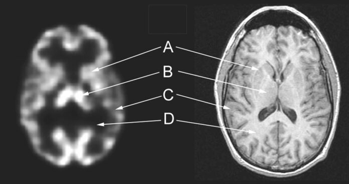
Brain SPECT imaging is a functional imaging technology that helps clinicians see how different regions of the brain are working in real time. Unlike traditional scans such as MRI or CT, which focus on the physical structure of the brain, SPECT visualizes blood flow patterns and metabolic activity. This paints a clearer picture of how the brain functions beneath the surface. It is especially valuable for diagnosing conditions that may not show up on structural scans, such as depression, ADHD, traumatic brain injury, dementia, and various neurological disorders.
At its core, brain SPECT imaging uses small amounts of radiotracers, which are safe pharmaceutical compounds that emit gamma rays, to highlight how well blood flows to different brain regions. Areas with healthy function show normal blood flow, while dysfunctional areas may appear cold (low activity) or hot (overactivity). These visual patterns give clinicians insight into how the brain is performing and help guide personalized treatment plans.
The Science Behind SPECT: A Journey Into Brain Function
Brain SPECT imaging, short for Single Photon Emission Computed Tomography, sits at the intersection of nuclear medicine and advanced diagnostic imaging. What makes SPECT unique is its ability to capture how the brain behaves rather than simply what it looks like.
The science behind SPECT centers on the concept of cerebral perfusion, or the movement of blood through the brain. Because blood flow is directly linked to function by providing oxygen, glucose, and essential nutrients, measuring perfusion allows clinicians to assess brain activity. Regions with healthy blood flow are typically functioning normally, while areas with reduced perfusion may indicate injury, inflammation, or degeneration.
Here is how the process unfolds scientifically:
- A radiotracer is introduced into the bloodstream.
- The tracer binds to specific receptors or distributes according to blood flow.
- As it moves through the brain, it emits gamma rays.
- A gamma camera rotates around the patient’s head to capture thousands of images.
- A computer compiles these images into 3D maps of brain activity.
This method allows clinicians to see not only which regions are active but also how different areas communicate and coordinate. Functional abnormalities such as underactive frontal lobes, overactive limbic regions, or uneven perfusion patterns become visible in ways that structural scans cannot reveal.
Ultimately, brain SPECT imaging offers a biological window into brain function, helping clinicians correlate symptoms with physiological findings and tailor treatment accordingly.
Decoding the Process: How SPECT Imaging Is Performed
A brain SPECT scan is a highly structured, step-by-step procedure designed to capture precise functional images.
1. Radiotracer Selection and Injection
The process begins with selecting a radiotracer based on the type of information needed. The most commonly used radiotracers for brain studies include:
- Technetium-99m HMPAO
- Technetium-99m ECD
- Iodine-123 based tracers
These substances are injected intravenously and circulate through the bloodstream. Once introduced, the tracers travel to the brain and settle in patterns that reflect blood flow and metabolic activity.
2. Resting Period for Absorption
After injection, patients typically rest quietly in a dim room for 30 to 60 minutes. This helps ensure that external stimuli do not influence brain activity, making the final images more accurate and reflective of the patient’s natural baseline.
3. Scan Acquisition
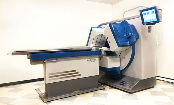
The patient lies comfortably on a padded table as the SPECT camera rotates around the head. During this time:
- The camera collects gamma-ray emissions.
- The patient must remain still to ensure high-quality images.
- Scanning usually lasts 20 to 60 minutes depending on the equipment and study protocol.
4. Image Reconstruction and Analysis
Advanced computer algorithms reconstruct the captured data into detailed, 3D functional maps. These images can be viewed from multiple angles and compared with clinical symptoms, cognitive testing, or other imaging studies. This comprehensive process provides a functional snapshot of how the brain is performing and helps identify areas that may require further evaluation or treatment.
Understanding Radioisotopes: The Key to SPECT Accuracy
Radioisotopes are the heart of brain SPECT imaging. Their unique properties make them ideal for capturing dynamic brain activity safely and effectively.
Why Radioisotopes Matter
Radioisotopes emit gamma rays, which are energy signatures that can be detected by SPECT cameras. Their distribution in the brain mirrors blood flow and metabolic processes, allowing clinicians to identify:
- Areas of reduced perfusion
- Hyperactive regions
- Inflammation or degeneration
- Functional abnormalities related to psychiatric or neurological conditions
Common Radioisotopes in Brain SPECT
Technetium-99m is the gold standard for brain SPECT imaging because:
- It has a short half-life of about 6 hours, which minimizes radiation exposure.
- Its gamma-ray emissions are optimal for high-resolution imaging.
- It binds reliably to brain tissue in proportion to blood flow.
Iodine-123 is another widely used isotope, often chosen when more specific biological pathways such as neurotransmitter activity need to be evaluated.
The Future of Radiotracers
As research evolves, scientists are developing new tracers that can target:
- Dopamine receptors
- Amyloid plaques associated with Alzheimer’s disease
- Inflammation markers
- Tumor metabolism
These innovations are expanding the diagnostic power of brain SPECT imaging and allowing physicians to understand complex conditions with greater precision than ever before.
The Transformative Benefits of Brain SPECT Imaging
Illuminating Mental Health: SPECT in Psychiatry
Brain SPECT imaging has transformed psychiatric care by revealing biological patterns behind mental health conditions. Disorders such as depression, anxiety, ADHD, bipolar disorder, and PTSD often show distinct cerebral blood flow patterns. Reduced activity in the prefrontal cortex, for example, is frequently seen in major depressive disorder. By providing measurable and visual evidence, SPECT helps clinicians tailor treatment plans more effectively and allows patients to better understand their own brain health.
Identifying Neurological Disorders: A Diagnostic Turning Point
Neurological conditions often remain hidden on traditional structural scans. Brain SPECT imaging detects functional disruptions, making it a powerful tool for diagnosing epilepsy, traumatic brain injury, dementia, Parkinson’s disease, and more. In epilepsy, SPECT can pinpoint the seizure focus. In dementia, SPECT distinguishes Alzheimer’s from other cognitive disorders by identifying characteristic blood flow reductions. These insights lead to faster, more accurate diagnoses and more effective treatment planning.
Personalized Treatment Plans Informed by SPECT Findings
One of the greatest strengths of brain SPECT imaging is its contribution to personalized medicine. By showing exactly how a patient’s brain is functioning, clinicians can develop individualized treatment strategies, whether adjusting medication, recommending neurofeedback, or targeting cognitive deficits. This reduces the guesswork often associated with psychiatric and neurological interventions and improves patient outcomes.
Exploring Clinical Applications: Where Brain SPECT Imaging Makes an Impact
Neurodegenerative Diseases: From Alzheimer’s to Frontotemporal Disorders
Early detection is essential for conditions like Alzheimer’s disease. Brain SPECT imaging allows clinicians to identify reduced blood flow patterns years before severe symptoms appear. It also helps distinguish between Alzheimer’s, vascular dementia, Lewy body dementia, and frontotemporal dementia. This functional clarity guides timely interventions, lifestyle modifications, and long-term planning.
Stroke Evaluation and Recovery
SPECT imaging helps determine the extent of stroke damage by mapping perfusion deficits. Clinicians can differentiate areas of permanent damage from regions that may be salvageable with immediate treatment. During recovery, SPECT can track improvements in blood flow, helping shape rehabilitation strategies that reflect the patient’s functional progress.
Applications Beyond Neurology
The usefulness of SPECT extends far beyond brain imaging. In cardiology, it assesses myocardial blood flow. In oncology, it helps evaluate tumor activity and treatment response. In rheumatology, it can assess inflammation in joints. These broad applications highlight the versatility and power of SPECT as a diagnostic tool.
Future Innovations: The Next Chapter in Brain SPECT Imaging

Advancements in Imaging Technology
Emerging detector technologies and improved reconstruction algorithms are enhancing SPECT clarity and resolution. Hybrid systems that combine SPECT and MRI provide both structural and functional insights in a single session. New radiotracers are also being developed to target specific neurotransmitter systems, opening doors to earlier detection of diseases like Parkinson’s long before physical symptoms appear.
Artificial Intelligence and SPECT
Artificial intelligence is beginning to reshape brain SPECT imaging by enhancing image interpretation, detecting subtle patterns, and predicting treatment outcomes. Machine learning can integrate SPECT data with clinical records to create more accurate diagnostic models. This technological advancement supports the move toward highly individualized care.
Expanding Global Access
Portable and cost-effective SPECT systems are being developed to bring advanced diagnostics to underserved regions. In areas with limited access to MRI or PET, brain SPECT imaging could become a crucial tool for diagnosing neurological and psychiatric conditions. As research evolves, SPECT may also support global health efforts in managing chronic illness, infectious disease, and brain health disparities.


